Flow Cytometry Core Facility
Multi-parameter measurement of characteristics of whole cells or cellular components
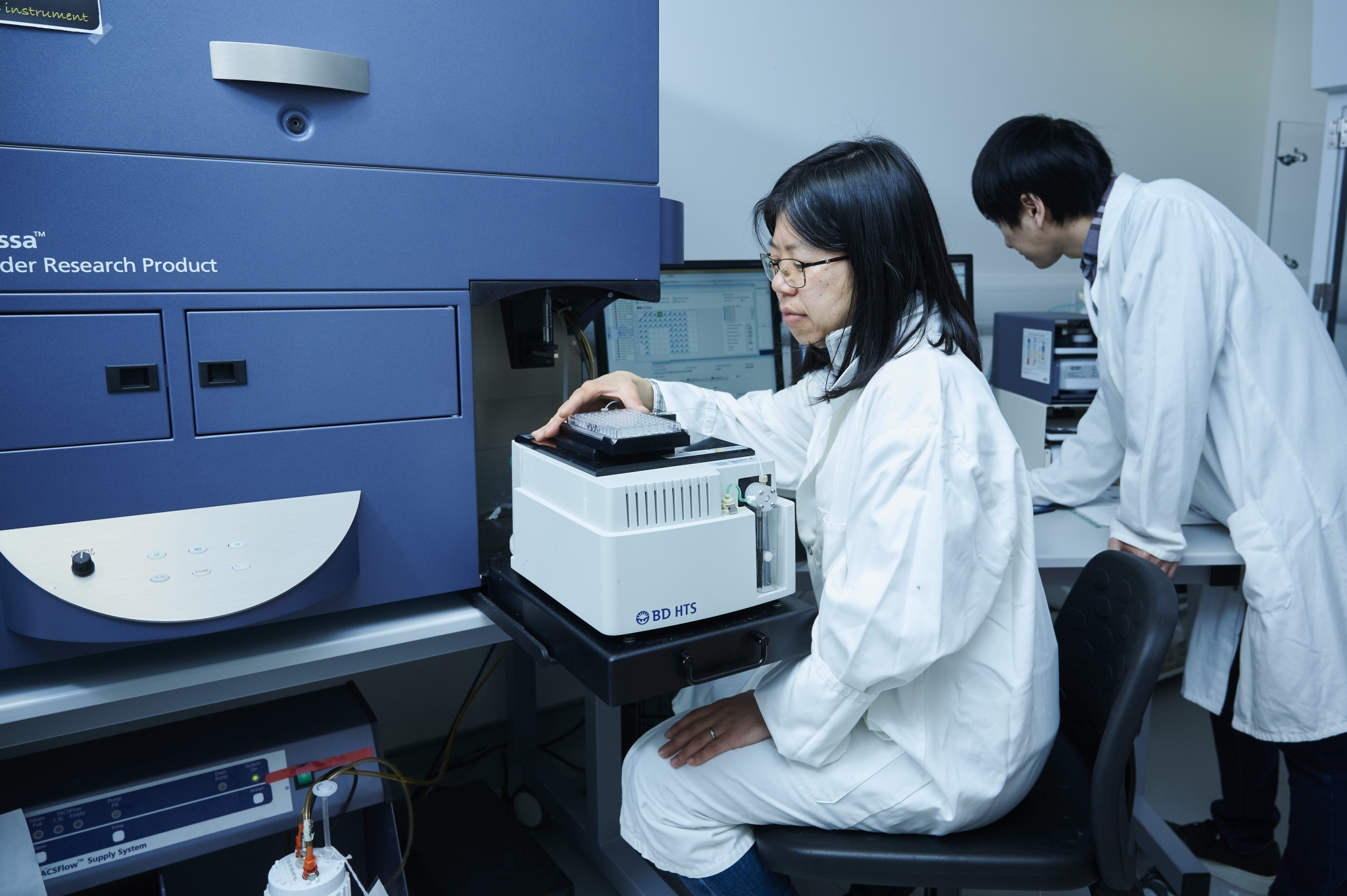
Researchers at the Flow Cytometry Core Facility
Maverick Photography
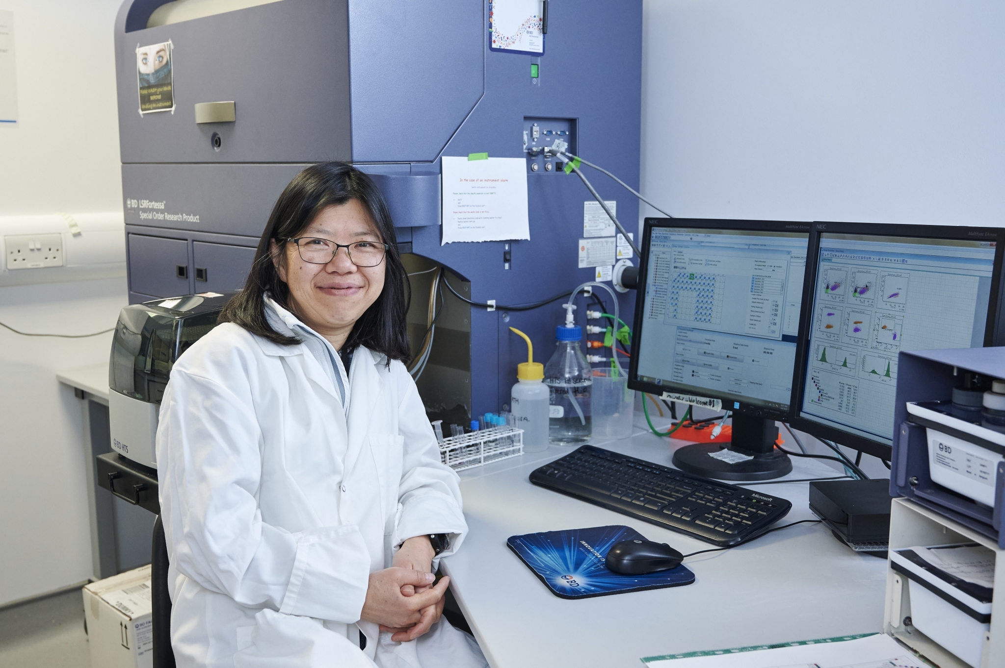
Dr Kumiko Samejima at the Flow Cytometry Core Facility
Maverick Photography
Qualitative and quantitative examination of whole cells and cellular constituents
Flow cytometry is a laser-based technology that allows the multi-parameter measurement of characteristics of biological particles in a single cell suspension as they flow in a liquid medium past an excitation light source.
Our Flow Cytometry faclity is intrumental for your biological research projects, providing qualitative and quantitative examination of whole cells and cellular components (such as nuclei or organelles).
Our expert staff are able to identify the biological particles as they flow in a liquid medium past an excitation light source, which strikes the individual moving particles, thus the light is scattered and fluorescence is emitted.
Industry sectors that can benefit:
- Agritech, agri-food and plant biology
- Healthcare and disease
- Life sciences and industrial biotechnology
- Pharmaceuticals and medical biotechnology
Facilities and expertise
We have 20 parameter multiplexing capabilities with aseptic cell sorting technologies, including 4-way simultaneously sorting and single-cell cloning. The facility also offers HTS plate acquisition with support for experimental design and analysis.
Equipment available
Benchtop analysers available for multiparameter analysis:
- FACSCalibur (BD) - dual laser - 488/633nm - 6 parameter
- FACSCanto II (BD) - 407/488/633nm - 2/4/2 colour
- LSR II (BD) - 355/407/488/633nm - 2/6/6/3 colour
- LSRFortessa (SORP-BD)- 407/488/561/633nm - 6/2/5/3 colour
Also available is a state-of-the-art high-speed cell sorter:
- FACSAria IIu Cell Sorter - 407/488/561/633nm - 6/2/5/3 colour
The FACSAria cell sorter provides a method for dissecting a heterogeneous mixture of biological cells into discreet sub-populations, one cell at a time, based upon the specific light scattering and fluorescent characteristics of each cell. It is a useful scientific instrument which provides fast, objective and quantitative recordings of fluorescent signals from individual cells as well as physical separation of cells of particular interest.
Case study: Studying mitotic chromosome structure using flow cytometry
Every organism is built from cells which are created through cell division. Genetic information, a manual encoding of how cells are built and maintained, is stored in the form of genomic DNA. Genomic DNA forms rod-shaped mitotic chromosomes during cell division which is necessary for the accurate transfer of genomic information. If genomic information is damaged or lost, the daughter cells could die or become harmful to the organism (e.g. give rise to cancer).
Dr Kumiko Samejima, a postdoc from the Earnshaw lab at the University of Edinburgh, was investigating how these mitotic chromosomes are formed using vertebrate cells as a model system. Previous studies showed that protein complexes called condensins are essential to forming rods. Kumiko has modified cell lines so that the condensin proteins can be rapidly depleted upon the addition of a chemical. She has further genetically modified these cells so they enter mitosis synchronously.
With these tools in place, Kumiko has been able to analyse the cell population entering mitosis minute-by-minute using inter-disciplinary approach: including high-resolution microscopy, genomic and proteomic analysis, and polymer modelling. Her data demonstrated that condensin organises DNA into loops arranged like a helical staircase along the axis of the cylinder.
The Flow Cytometry Core Facility has been integral to these studies since obtaining a cell population uniformly depleted of target protein (e.g. condensin) is critical to generating reliable experimental data. Kumiko used flow cytometry to measure the levels of each fluorescently-labelled target protein in individual cells. This is essential to establish and maintain cell lines that exhibit maximal and even levels of depletion of target proteins. Cell sorting is also crucial to eliminate those cells with high levels of target proteins that have managed to evade the depletion procedure.
Kumiko collaborated with Facility Manager, Martin Waterfall, to establish a new protocol to synchronise cells, obtaining a SULSA technology seed project grant to develop a method obtain cells in the later stages of mitosis termed anaphase/telophase. It is hoped this new method will become a valuable tool for understanding how genomic DNA is transferred safely into new cells.
Find out more
Visit the website for the Flow Cytometry Core Facility
Interested in learning more?
Speak to our expert

Cameron Chalmers
Consultancy Manager

Cameron Chalmers bio
Cameron Chalmers
Consultancy Manager