The Cryo-Electron Microscope Facility
High-resolution imaging of biological samples under cryogenic conditions as well as electron crystallography capability.
Maverick Photography
Industry sectors that can benefit from these facilites:
- Agri-tech, agri-food and plant biology
- Food and drink
- Healthcare and disease
- Life sciences and industrial biotechnology
- Pharmaceuticals and medical biotechnology
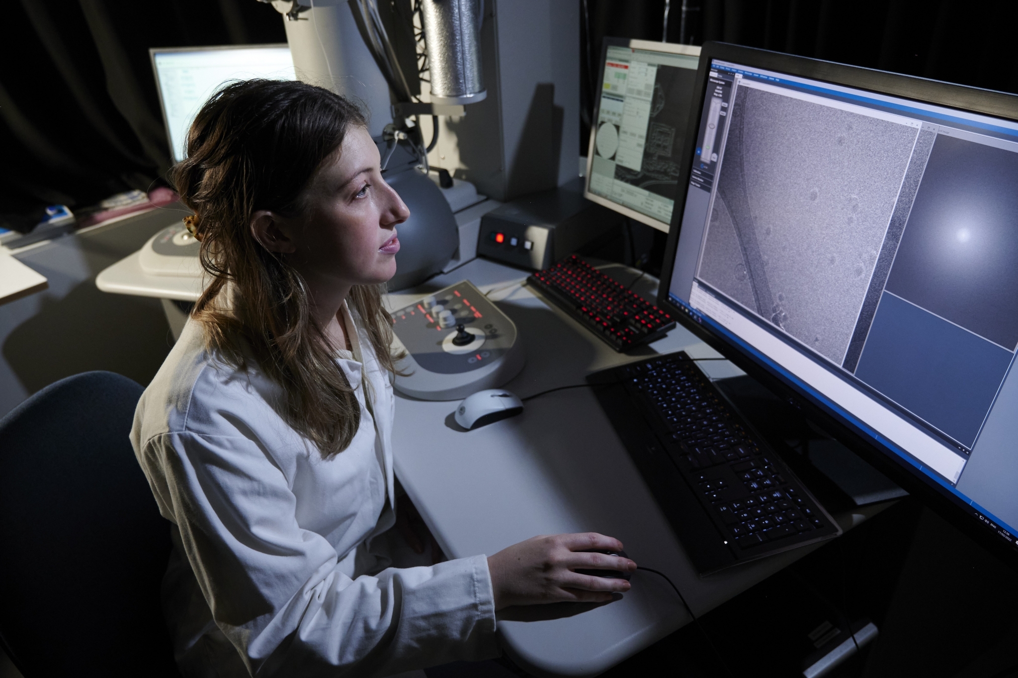
The Cryo-EM facility offers high-resolution imaging of biological samples under cryogenic conditions as well as electron crystallography capability.
Techniques:
- Standard 2D projection imaging of thin samples.
- Single-particle analysis of proteins is the core strength of the facility.
- (Cryo-) electron tomography provides 3D information of a single specimen by physically tilting the specimen inside the microscope.
- Electron diffraction provides structural information on crystalline material.
- Extensive training (or service) for users, ranging from negative stain sample preparation, conventional room-temperature TEM, to plunge-freezing, cryo-TEM and processing of single-particle data.
Equipment available:
- Thermo Fisher Scientific Tecnai F20 electron microscope (200 kV, field emission gun) equipped with a 3k x 3k CMOS camera (Gatan Rio) and Gatan K2 direct electron detector.
- Gatan single tilt liquid nitrogen cryo-transfer holders (Gatan 626 and 698), with transfer-stations, pumping station and temperature control units.
- Thermo Fisher Scientific Vitrobot automated vitrification unit (Vitrobot mark IV).
- Various supporting equipment, such as a glow-discharge unit, negative stain facilities and a room-temperature holder.
- Extensive computer resources for data processing.
More details
Contact Martin Singleton, Facility Manager
Find out more about the Cryo-Electron Microscope Facility
Maverick Photography
Example use of facilities:
Helping researchers to see how pathogens communicate
The University of Edinburgh has world-leading expertise in understanding pathogens and how they communicate with host cells to establish infections and transmit between individuals.
Dr Amy Buck’s lab investigates how pathogens release small extracellular vesicles to shuttle their molecules directly into cells in order to change the immune response. However, vesicles are tiny (100-1000 fold smaller than a human cell).
PhD student Ruby White used the Cryo-EM facility which enabled her to visualise the native structures of the vesicles and understand the surface molecules that enable them to enter specific host cells.
This basic research not only informs the tricks that pathogens use to survive in their hosts, but could also lead to basic understanding of vesicle trafficking that is relevant to other diseases. For example, vesicles are important in neurodegeneration and cancer, where they are released from tumours and can change cells in the microenvironment.
Interested in learning more?
Speak to our expert

Michelle Holland
Head of Operations
Michelle Holland bio
Michelle Holland
Head of Operations
Consultancy & Commercial Services